 |
A 42yo man is admitted to ICU through the emergency department, having presented with central abdominal pain, nausea and vomiting. The patient denies alcohol ingestion or illicit drug use. He is clammy, with the following physiology:HR: 112bpm SR BP: 114/62mmHg RR: 32bpm SpO2: 97% on O2 10L/min via facemask Temp: 37.8C |
On examination, he has decreased breath sounds in both bases with scant inspiratory crepitations and his abdomen is tender in the epigastrium and periumbilical regions with mild guarding.
A point-of-care ABG shows the following result:
|
pH 7.22 |
 |
The emergency department consultant has recently completed a Certificate of Physician Performed Ultrasound and has performed a bedside abdominal ultrasound that demonstrated a normal calibre aorta, a normal gallbladder and two normal kidneys.
Describe and interpret the results from the point-of-care ABG.
A raised anion gap (16) moderate metabolic acidosis with an appropriate compensatory fall in the PaCO2. The lactate is not elevated, suggesting an alternative reason for the elevated anion gap. The patient denies alcohol ingestion, leaving ketones, azotaemia and medications. Also, the HCO3- value is not fully explained by the raised anion gap component, suggesting a co-existing normal anion gap metabolic acidosis, most likely due to associated renal impairment or lower GI losses.
The hyperglycaemia suggests DKA or HHNS, with DKA the more likely given the degree of acidosis and the corrected Na+ of 138mmol/L. The calculated serum osmolarity is 285mOsm/kg before adding the urea (value not given), suggesting the patient is likely to be hypersomolar, unless the urea is low.
Mild hypokalaemia, but in the setting of a likely DKA, this is likely to reflect depleted total body stores.
Some of the biochemistry results return as follows:
Urea 12.1mmol/L
Creatinine 114umol/L
Na+ 131mmol/L
K+ 3.1mmol/L
Cl- 103mmol/L
HCO3- 14mmol/L
Amylase 76U/L
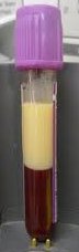
Comment: “White layer noted on top of the blood sample. Further testing awaited.”
What diagnosis does this information suggest? What test do you think is being performed and what cut-off value would be of concern? What are the treatment options for this particular aetiology?
A white layer forming at the top of a blood sample in this context suggests hyperlipaemia, though severe leucocytosis as occurs with leukaemias can give a similar appearance. Given the absence of a TPN or propofol infusion, in this patient it is likely to represent hyperlipidaemia, specifically hypertriglyceridaemia in this patient. The likely diagnosis therefore, is hypertriglyceridaemic induced acute pancreatitis, precipitating a diabetic ketoacidosis.
The test being performed is a triglyceride level and values above 5.6mmol/L (500mg/dL) are consistent with a severe hypertriglyceridaemia, though levels are usually much higher. The absolute TG level does not correlate with the severity of the pancreatitis.
Hypertriglyceridaemia is reported to account for up to 38% of cases of acute pancreatitis in general and is the third most common cause of non-iatrogenic pancreatitis after gallstones and alcohol. Up to 56% of pancreatitis in pregnancy is related to hypertriglyceridaemia (Gallstones account for about 35%.)
Hypertriglyceridaemia can interfere with the amylase assay resulting in a false negative in the setting of acute pancreatitis. (Yes, most centres perform lipase asssays. I’m just making a point.)
A 1995 paper in the American Journal of Gastroenterology described 3 populations associated with hypertriglyceride induced pancreatitis
- Poorly controlled diabetics with hypertriglyceridaemia
- Alcohol abusers with hypertriglyceridaemia
- Non-alcoholic, non-diabetic, non-obese patients with diet or drug induced hypertriglyceridaemia.
Conditions other than diabetes, alcoholism and obesity that are associated with hypertriglyceridaemia include pregnancy, nephrotic syndrome, hypothyroidism, certain medications such as propranolol, anti-protease antiretroviral drugs, oestrogens, olanzipine, mirtazipine and retinoids. Hypertriglyceridaemia may also be primary, as in all but the class IIa Fredrickson classification of inherited hyperlipidaemias.
For TG levels less that 5.6mmol/L, treatment consists of dietary modification and exercise. Statins are usually the first line medications as the TG levels are often not raised in isolation from the other lipid fractions. However the fibrates are more effective at lowering TG levels and are preferred when TG levels are >5.6mmol/L. Fibrates appear to impair statin metabolism and when combined, the risk of statin induced rhabdomyolysis may be increased.
For severe hypertriglyceridaemia, with or without pancreatitis, several additional therapies have been advocated, the the evidence quality is low, consisting mainly of case series and small single centre trials. These therapies include apheresis, insulin (subcutaneous or by infusion) and heparin. The therapeutic target is reduction of the TG level to <5mmol/L.
References
- Hyperlipidemic pancreatitis. Toskes PP. Gastroenterol Clin North Am. 1990;19(4):783.
- http://www.gpnotebook.co.uk/simplepage.cfm?ID=-194314232&linkID=34086&cook=no&mentor=1
- http://emedicine.medscape.com/article/126568-overview
- Pancreatitis may occur with a normal amylase concentration in hypertriglyceridaemia. Sharma P, Lim S, James D, Orchard RT, Horne M, Seymour CA. BMJ. 1996;313(7067):1265.
- Thompson PD, Clarkson P, Karas RH. Statin-associated myopathy. JAMA. 2003;289:1681–90.
- Management of acute severe hyperlipidemic pancreatitis. Kyriakidis AV, Raitsiou B, Sakagianni A, Harisopoulou V, Pyrgioti M, Panagopoulou A, Vasilakis N, Lambropoulos S. Digestion. 2006;73(4):259.
- Treatment of severe hypertriglyceridemia in nondiabetic patients with insulin. Mikhail N, Trivedi K, Page C, Wali S, Cope D. Am J Emerg Med. 2005;23(3):415.


