
By Oli Flower
These are images from real ICU patients. The stories and details are changed to preserve confidentiality.
[su_tabs][su_tab title=”History”]
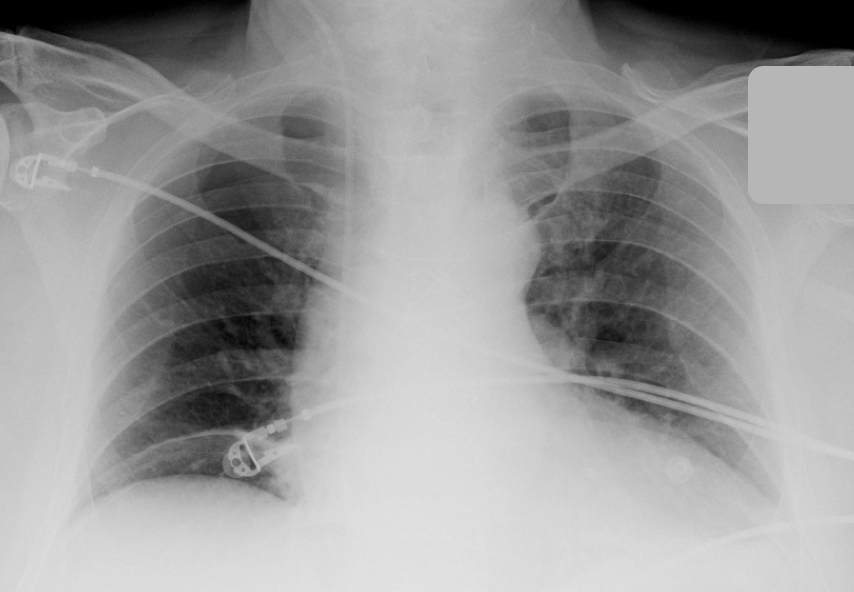
70 year old man 2 days after cardiothoracic surgery
His chest drains were removed earlier in the day
An AP semi erect chest X-ray is taken
[/su_tab][su_tab title=”Image”]
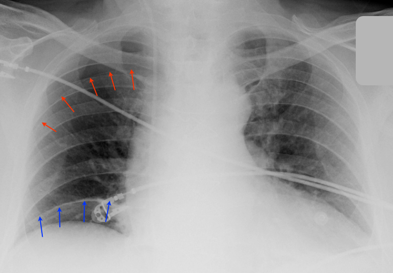
The blue arrows show a band of linear atlectasis that at first glance gives the appearance of air under the diaphragm
This was not the case as became evident in subsequent films
This distracts from a more important diagnosis: a pneumothorax (red arrows)
There was no respiratory compromise and the pneumothorax was managed conservatively
[/su_tab][su_tab title=”More Resources”]
British Thoracic Society Guidelines on Management of Pneumothorax
Lots of great pneumothorax images
A video showing one approach to inserting a formal chest drain for a pneumothorax
A video showing a Seldinger technique for insertion of a chest drain
[/su_tab]
[/su_tabs]


