
Case 22 from Drew Sullivan
[az_accordion_section] [accordion title=”History” id=”acc-1″]A 45 year old man presents with a 2 week history of diplopia. His golfing friends had noticed that his game was ‘off’ and he was frequently and uncharacteristically ‘swinging and missing’. On examination, you observe right ptosis, mydriasis and a ‘down and out’ pupil. You proceed to CT:[/accordion] [accordion title=”Non-contrast CT Brain” id=”acc-2″]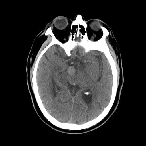
 What abnormality is present? Which investigation would you order next?[/accordion] [accordion title=”CT Cerebral Angiography” id=”acc-3″]Axial CT Cerebral Angiogram
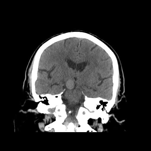
3D Reconstruction[/accordion] [accordion title=”Answer” id=”acc-4″]The non-contrast images demonstrate a well-defined round, slightly hyper-attenuated structure in the prepontine and interpeduncular cisterns. No acute haemorrhage is identified.
The CT Cerebral Angiogram and 3D Reconstruction confirmed a 12 x 18mm basilar tip aneurysm. Incidentally, the left posterior cerebral artery has a fetal origin (supplied by left posterior communicating artery) – a normal variant.
The patient’s signs and symptoms are of a unilateral third nerve palsy. The pupil is ‘down and out’ because of the unopposed actions of lateral rectus (CN VI – Abducens nerve) and superior oblique (CN IV – Trochlear nerve). The remainder of the extra-ocular muscles are supplied by CN III (oculomotor nerve) which is compressed by the aneurysm as it emerges from the midbrain.
The patient proceeded to have elective craniotomy and clipping of the aneurysm.
The 5 year cumulative risk of rupture of posterior circulation aneurysms:
<7mm: 2.5%
7-12mm: 14.5%
13-24%: 18.4%
>25mm: 50%[/accordion] [/az_accordion_section]


