• Twitter
  • Facebook
  • Vimeo
  • FCUS Courses
  • Register for FCUS Course
  • Login
Intensive Care Network
  • About Us
    • What is the ICN?
    • The ICN Story
  • Events
  • Podcast Series
    • ICN Activities
      • ICN Hot cases
      • Semantic sMatter
      • ECG Proving Ground
      • Echo Cases
      • SMACC 2013
      • ICU Radiology Cases
      • Game Changing Evidence
  • Regional Networks
    • SIN
    • BIN
  • Exams
    • Primary ANZCA
      • Pharmacology
      • Physiology
    • Primary ACEM
      • Pharmacology
      • Physiology
      • Pathology
      • Anatomy
    • Fellowship CICM
      • Notes
      • Past Papers
    • Fellowship Paeds
      • Approaches to Questions
      • Clinical Exam
      • Core ICU
      • Clinical Governance
      • Medicine
      • Surgery
    • Fellowship ANZCA
      • Notes
      • Past Papers with Answers
  • Resources
    • Links
    • The ICN meta feed
    • Reviews
      • Courses
      • Conferences
      • Websites & Blogs
      • Books & Journals
    • Clinical Resources
      • Clinical Calculators
    • Non-clinical resources
      • Research
  • Discussion Forum
    • Main Page

Radiology Case 23

Home Radiology Case 23

Case 23 from Drew Sullivan

[az_accordion_section] [accordion title=”History” id=”acc-1″]A 70 year old man presents to the Emergency Department following a witnessed syncope whilst seated. He describes a 2 day history of intermittent palpitations with associated light-headedness but no chest pain or shortness of breath. This is his first episode of syncope. His examination findings were benign. You proceed to order a chest radiograph.[/accordion]

[accordion title=”CXR” id=”acc-2″]Case 23 CXR Unlabelled
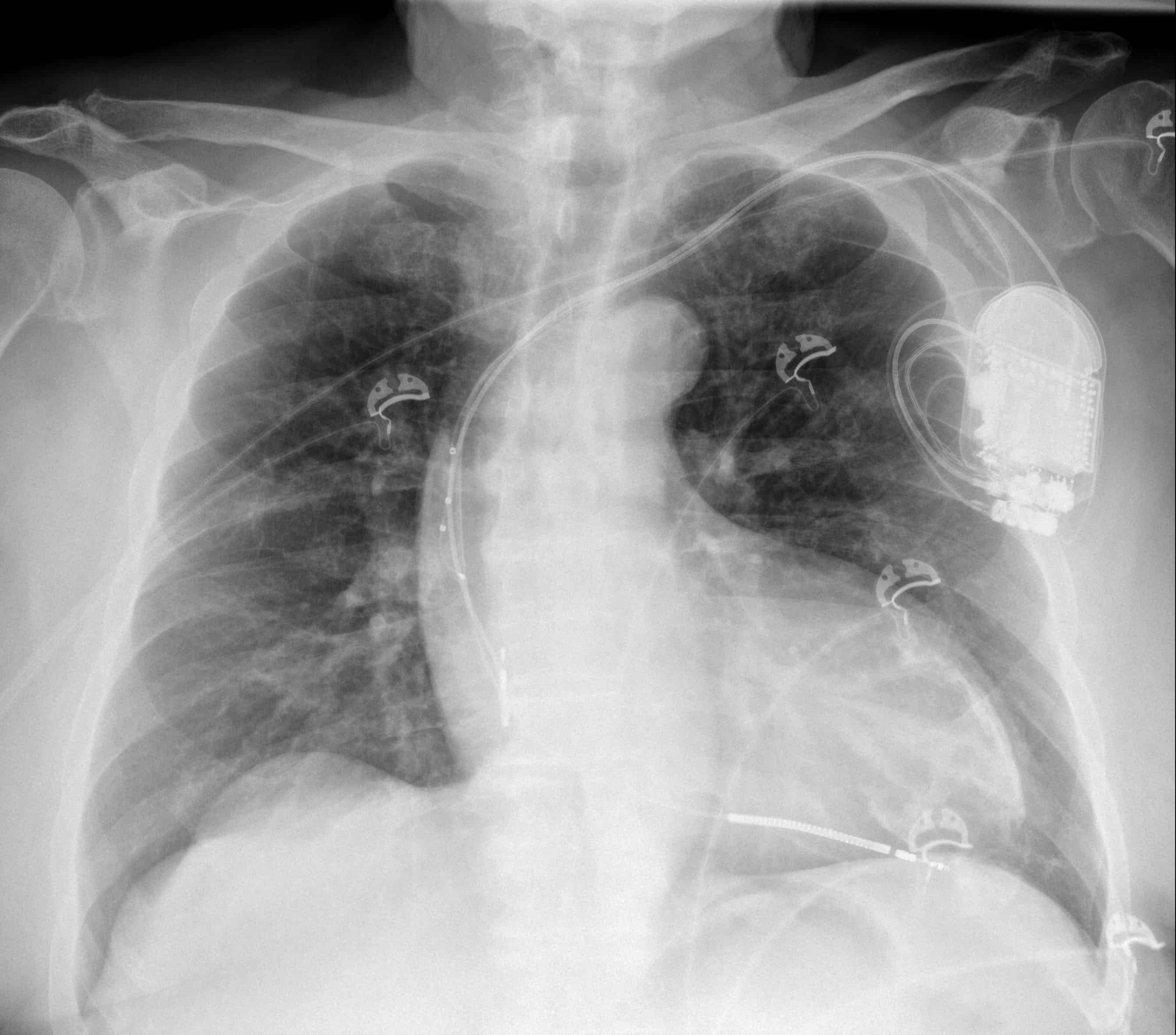
What are the two most significant findings on this radiograph?[/accordion]

[accordion title=”Answer” id=”acc-3″]Case 23 CXR Labelled

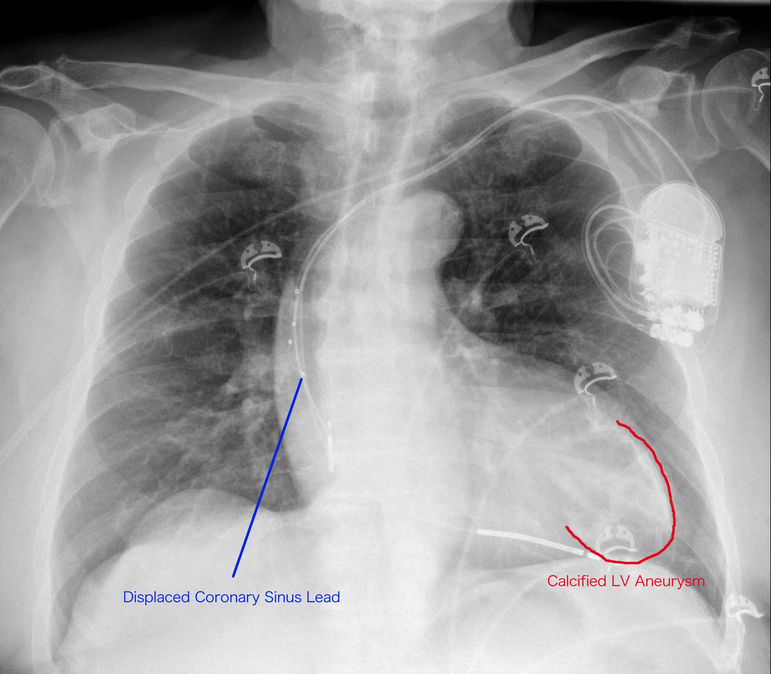
There is an AICD in-situ.  The right atrial and right ventricular leads are in satisfactory position, however the coronary sinus lead has migrated back into the superior vena cava.

There is also coarse curvilinear calcification projected over the left ventricle consistent with a calcified left ventricular aneurysm, a consequence of a previous myocardial infarction.

Compare this examination to the radiograph performed post-AICD insertion in which the lead positions are appropriate.
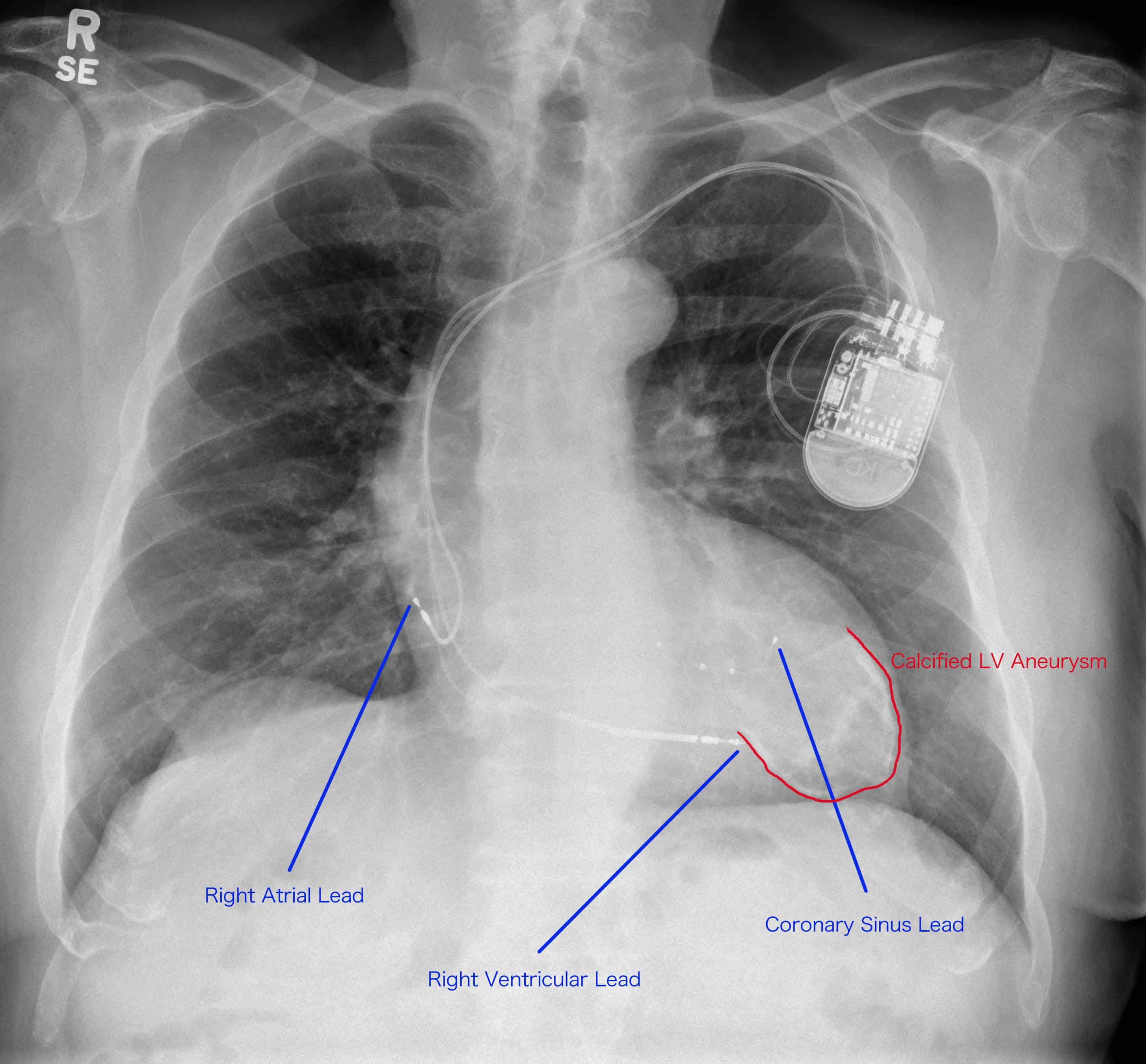
Case 23 CXR Labelled 2[/accordion]

[accordion title=”Resources” id=”acc-4″]Radiographics (RSNA) – Radiography of Cardiac Conduction Devices:  A Comprehensive Review[/accordion][/az_accordion_section]

Share this
FLASHBACK FRIDAYFLASHBACK FRIDAY - BURNS
IPSN – Intensivist Parent Support Network WIN – Women in Intensive Care Network ICN UK ICN NZ ICN WA ICN VIC ICN NSW ICN QLD The ICN Story The Team Jellybean Podcasts ICN Blog SMACC Video SMACC Audio Video ECG Simulator by aclsmedicaltraining BASIC SCIENCE CLINIC Simulation Game Changing Evidence ICU Radiology Echo Guide ECHO Cases Clinical Cases EXAMHELP Jellybean Podcasts ICN Blog SMACC Video SMACC Audio Video CICM Fellowship ANZCA FELLOWSHIP PAEDS FELLOWSHIP EMERGENCY PRIMARY ANAESTHETICS PRIMARY End-o-bed-o-gram
  • About
    •  What is ICN
    •  The Team
    •  ICN NSW
    •  ICN QLD
    •  ICN VIC
    •  ICN WA
    •  ICN NZ
    •  ICN UK
  • Resources
    •  Lung US
    •  Exam Help
    •  Clinical Cases
    •  Echo Cases
    •  Echo Guide
    •  ICU Radiology
    •  Game Changing Evidence
    •  ICN Metafeed
    •  Simulation Resouces
  • Media
    •  SMACC Posters
    •  Audio
    •  Video
    •  Pecha Kuchas
  • Upskill
    •  Clinical Cases
    •  Echo Cases
    •  ICU Radiology
  • Exam Help
    •  End-o-Bed-o-Gram
    •  ICU Primary Exam
    •  CICM Fellowship
    •  ANZCA Fellowship
    •  Paeds Fellowship
    •  Emergency Primary
    •  Anaesthetics Primary

® 2026 The Intensive Care Network || All rights reserved || Disclaimer || Site Map || Contact ICN Support

Log in with your credentials

or     Create an account

Lost your password?

Forgot your details?

I remember my details

Create Account